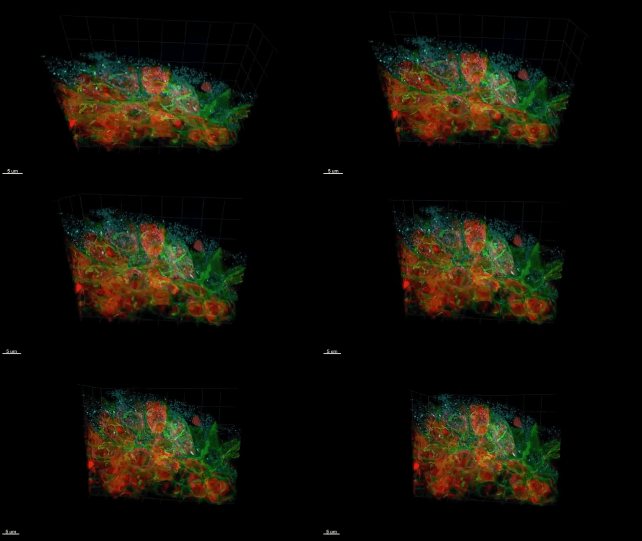
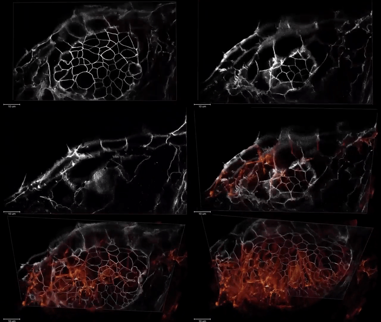
Group leader Eric Betzig and his team of researchers at HHMI Howard Hughes Medical Institute have created up an amazing microscope that can view deep, subcellular movement within multi-cellular organisms and captures high resolution, dynamic 3D images and footage of said movement. By combining features such as “sheer lattice microscopy” with “adaptive optics”, the team has been able to see what could never be seen before. Such example of this footage is shown in the immune cell migration in the zebrafish inner ear, a remarkable witness to how immune cells develop within zebrafish embryos.
The new microscope is essentially three microscopes in one: an adaptive optical system to maintain the thin illumination of a lattice light sheet as it penetrates within an organism, and another adaptive optical system to create distortion-free images when looking down on the illuminated plane from above.
Another such example of this sub-cellular footage is Endocytosis in a human stem cell derived organoid..
This microscope also captured the formation of membrane dynamics in the zebrafish eye“.
More of this fascinating footage is available through the HHMI site.
via Digg


